
SCANNING PROTOCOL: Rx Best-Quality
ACTUAL DOSE: 10.00 mA x 75.00 Kw
CLINICAL INFORMATION: Rx OPG scan of first clinical investigation, in a Patient suffering from irritating or blunt pain in the throat, head, neck and face. This pain has persisted for approx. 2 months and is resistant to drug therapy.
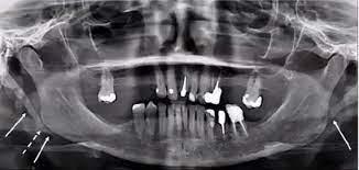
RADIOLOGICAL ANALYSIS
- The cortical bone is found to be within physiological parameters.
- There are no pathological alterations to the Patient’s medullary trabecular structure.
- Both condyles and glenoid fossae appear of normal radio morphology.
- There is evidence of periodontal disease, with consequent perialveolar bone resorption.
- Presence of mild lesions due to osteoporosis.
- Asymmetrical calcification of the SH Process is present, providing an indication for the diagnosys of Eagle’s Syndrome. Clinical confirmation is due.
IN-DEPTH…
Introduced in 1937 by American otolaryngologist Watt Weems Eagle, this syndrome, named after the aforementioned physician, is a pathology of the SH process.
The styloid apophysis is a thin bony process located at the base of the temporal bone, right behind the mastoid apex: it has anteromedial and caudal placement to the maxillo-vertebro-pharyngeal cavity, which contains carotid arteries, internal jugular vein, facial nerve, glossopharyngeal nerve, vagus nerve and hypoglossal nerve; At this point come in the stylohyoid ligament, which is a fibrous fascia connecting the styloid with the hyoid. The latter flanks the stylohyoid muscle, which grafts on the apophysis itself, and together with the stylo-glossus and the stylo-pharyngeus muscle, helps to control mouth’s floor, tongue and larynx.

PATHOLOGY
The Styloidic or Eagle’s syndrome is a disorder of the ligaments connecting the skull to the hyoid bone in the neck, which after calcification, end up causing painful symptoms in the mouth, head, neck, throat and forehead. Particularly, the syndrome is caused by the lengthening of the styloid process, a small bone at the base of the skull, and/or by calcification of the ligament. This condition typically occurs after an injury or trauma to the neck or pharynx, or after surgery in the neck region, such as tonsillectomy.
CAUSES
Its exact causes are not yet known, although repeated trauma to the neck, irritation or scarring of this region, muscle spasms (as in the case of cricopharyngeal spasm) and dyskinesias of the hyoid or sublingual muscles appear to promote the disorder: stiffening of the ligament, which may end up suffering from ostosization phenomena, is the underlying etiopathogenetic element of all symptoms; the higher incidence in women over 40 years and a certain association with senescence suggests a relationship with menopause and endocrine disorders.
Some researchers suspect that the syndrome may result from trauma caused in the developmental stages of the styloid process; a trigger can be local trauma or surgical stress, such as a tonsillectomy triggering chronic dull pain on the affected side of the head, sometimes accompanied by swelling between the roof of the mouth and the back of the throat on the same side.
In other cases, deformation of the styloid process extends laterally, compressing the internal or external carotid artery, with pain radiating along the artery to one side of the neck when the head is turned, pain above or below the eye, depending on which artery is involved: this form is called stylo-carotid.
Tensions existing in the sub-occipital area and in the chewing area may have a key role in the genesis of the SH syndrome, as the styloid is inserted in the kinematic chain connecting the head with the shoulders through the hyoid bone: maintenance of correct neuro-myofascial ratios between styloid, mastoid and mandible apophysis allows to guarantee centrality of the body and correct positioning of the skull, and consequently, of the Frankfurt plane ground-wise and horizotally, regardless of any actions performed, such as breathing, speaking, and swallowing.
SYMPTOMS
Manifestations of Eagle’s syndrome include:
- Pain persisting for days, weeks, or even months
- Ear pain on the same side of pain in the head, neck, forehead
- Feeling of something stuck in the throat
- Pain when swallowing (Odinophagy)
- Hoarseness of the voice (Acute Dysphonia)
- Excessive salivation
- Other symptoms may include: cough, dizziness, migraine, occipital neuralgia, tooth and jaw pain, sinusitis or bloodshot eyes in severe cases.
DIAGNOSIS
Lainglais classification based on OPG x-ray imaging. Bears little to no use nowadays, apart from observation of the syndrome, which requires diagnostic investigations to be confirmed. In all cases of chronic facial pain, such a finding deserves further investigations in the presence of significant clinical signs and symptoms.
Diagnostic Investigations involve Functional Tests such as:
- CT / CT angiography
- FMR (Functional Magnetic Resonance)
- ECOCOLOR DOPPLER
- TRANSORAL ULTRASOUND
- INTRAVASAL ULTRASOUND
Elaborated by Dr. Lucisano Francesco, Lead Radiographer – DentQ Italy